MEET THE SURGEON

Kevin J. Malone, MD
Associate Professor
Case Western Reserve University School of Medicine
Department of Orthopaedic Surgery
Chief of Hand and Upper Extremity Surgery
University Hospitals Cleveland Medical Centre
OVERVIEW
A 17-year-old right-hand-dominant male presented with a flexed, swollen and painful right ring finger ten days following an injury sustained when his hand got caught in a dog leash.
X-ray revealed a PIP joint fracture, involving a large P2 volar lip fragment and comminution from the base of the middle phalanx. Dr Malone achieved definitive fixation using a volar approach with two 1.5mm cannulated Micro Screws.
CASE INTRODUCTION
A 17-year-old male student, presented with an injury to the right ring finger of his dominant hand after a dog leash wrapped around it. Ten days after the incident, he sought medical attention as theinjury failed to resolve. Imaging showed a PIP joint fracture involving the base of the middle phalanx, with a large P2 volar lip fragment.
CASE PRESENTATION
The patient presented with acute onset pain, swelling and reduced range of motion (ROM) in his right ring finger after trauma. As is commonly seen, the patient initially ignored the injury in the hope it would resolve, however after ten days he sought medical attention due to persistence.
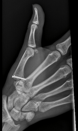
On examination the finger presented flexed and inflamed in the vicinity of the PIP joint, with reduced range and pain on motion. Pre-operative imaging (shown below) revealed a comminuted displaced intra-articular fracture at the base of the ring finger middle phalanx without apparent PIP joint subluxation.
Below: Pre-operative Imaging


PRE-OPERATIVE PLAN
Given the patient’s age, profession, leisure activities, handedness and delayed presentation, Dr Malone opted for open reduction internal fixation (ORIF). Due to the propensity for scarring and long-term stiffness, Dr Malone planned to use a volar approach with definitive fixation using lag screws due to the potential to reduce short term morbidity and the strong correlation between early active motion and improved outcomes.
Dr Malone selected the 1.5mm cannulated Micro Screw, as the implant size is highly appropriate for the management of phalanx trauma and is purposefully designed for small and fragile fragments. In addition, the cannulated design of the 1.5mm Micro Screw provides an unprecedented advantage over traditional small diameter screw options. This cannulation was considered to be required for this case to simplify the precise management of the volar lip fragment.
On reflection, Dr Malone notes that whilst traditional teachings suggested location of the joint is more important than concentricity, recent authors have noted a higher correlation of post arthritis if the joint is not anatomically reduced. The Micro Screw facilitates less soft tissue disruption, earlier return to function and a potential reduction in post-operative issues.
Dr Malone rejected the use of a dorsal splint due to concerns of stiffness and morbidity. He noted a key benefit of the Micro Screw was its suitability for a volar approach and assistance with improved reduction, which allowed for a shorter operation and quicker return to function.
SURGICAL APPROACH
Dr Malone used a volar approach, electing to perform ORIF of the right ring finger middle phalanx to avoid the dorsal structures which are prone to stiffness.
Reduction
Dr Malone made a volar Bruner incision centered at the PIP joint of the right ring finger and dissected down to the flexor tendon sheath while elevating flaps medially and laterally. The radial and ulnar neurovascular bundles were identified, mobilised and protected. The flexor tendon sheath was opened along the distal margin of the A2 pulley and extended out to the proximal margin of the A4 pulley. A quarter inch Penrose drain was placed around the FDP and FDS tendons within the tendon sheath to facilitate mobilisation of the structures away from the volar plate of the PIP joint. The volar plate, which was connected to the displaced volar rim fragment, was identified. A longitudinal incision along the radial and ulnar margins of the volar plate back towards the proximal
phalanx neck was made which allowed rotation of the volar fragment on its volar plate pedicle. This exposed the articular surface of the proximal phalanx head and middle phalanx base. With distraction, it could be seen that a large portion of the central articular surface was affected. The fracture line was very carefully re-established so that the articular surface could be elevated back to its anatomic position.
Bone preparation
Once reduction was confirmed with fluoroscopy, the fracture was stablised with a 0.011-inch K-wire retrieved dorsally along the base of the middle phalanx and drawn all the way into the fragment, to ensure it was not prominent. This was confirmed with fluoroscopic imaging. The volar rim piece on its volar plate pedicle was brought back into anatomic position and secured. A 0.011 inch guidewire was advanced from the radial aspect of the space across the subchondral bone of the middle phalanx, directed towards the central slip insertion.
Definitive fixation
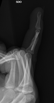
Dr Malone measured and selected an 11mm x 1.5mm cannulated Micro Screw from Field Orthopaedics. The volar segment was over-drilled and the Micro Screw inserted over the guidewire. Once fully seated, fluoroscopic imaging confirmed no evidence of dorsal cortical penetration and both guidewires were removed. In a similar fashion, a 9mm x 1.5mm Micro Screw was implanted along the ulnar aspect of the fracture for additional stability. Anatomic alignment and implant position were confirmed by fluoroscopic imaging (see images below).
Below: Intra-operative fluoroscopic images


Closure
The collateral ligaments were left preserved for good stability and there was no evidence of subluxation or instability when the PIP joint was taken through full ROM. The wound was irrigated, incisions repaired and the drain removed. The flexor tendon was allowed to slide back into the anatomic position and the flexor tendon sheath repaired to provide a good gliding surface. The wound was closed in interrupted fashion. The patient was placed in a well-padded ulnar gutter splint holding the ulnar digits into an intrinsic plus position.
FOLLOW UP
At follow up, the patient reported an absence of any pain in his right hand and despite a 5-10 degree flexion contracture, common with these injuries, the patient had re-established full composite digital ROM and grip strength. Further examination revealed a completely healed surgical incision in the palmer aspect of the right ring finger, hyperextension of all DIP joints and no tenderness
around the PIP joint. Dr Malone considered this to be an outstanding outcome.
Below: Six weeks post-operative ROM


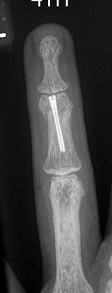
Below: Six weeks post-operative x-rays


DISCUSSION
In this case, Dr Malone was able to effectively address, in the short-term, an injury pattern that inflicts a high morbidity and often has long lasting notable consequences. It is considered that with other treatment approaches, either non-operative or operative, there is a requirement for more extensive follow up and active motion would have been delayed. The key benefits of the Micro Screw in this application were the ability to provide a definitive operative solution through a minimal incision, the small implant size provided ridged fixation even for the small fragment (as seen with the volar lip) and the impressive implant strength allowed for a quicker return to function (reducing morbidity and providing a significant saving on follow up costs and time).
The PIP joint is a key structure of the hand with fracture being a very common occurrence that can be complicated to manage, traditionally leaving patients suffering permanent stiffness and prolonged morbidity. This case highlights the Micro Screw as a highly beneficial modern solution which, when combined with an evidence-based surgical technique, was able to address a number of common contributors to complication and reliably return the patient to normal function.