MEET THE SURGEON

Timothy B. Larson, MD
Orthopaedic surgeon at Denton Hand & Orthopaedics,
Denton Texas
Special interest in advanced and minimally-invasive
surgical techniques for repair of the hand and wrist.
Fellow of the Mary S. Stern Hand & Upper Extremity
Fellowship program, Cincinnati Ohio.
DISCLAIMER
The opinions expressed by Dr. Larson are those of Dr. Larson and not necessarily those of Field Orthopaedics. Individual experiences may vary.
The procedure described in the report may differ from the manufacturer’s surgical technique. Surgeons are advised to review the product specific surgical technique prior to performing any surgery.
A surgeon must always rely on their own professional clinical judgement when deciding whether to use a particular product when treating a particular patient. A surgeon must always refer to the instructions for use, product label and surgical technique before using any Field Orthopaedics product. Product may not be available in all markets. Please contact your Field Orthopaedics representative if you have any questions about availability of Field Orthopaedics products in your area.
OVERVIEW
A 20-year-old right-hand dominant male presented to Dr Larson complaining of thumb pain, three days after falling off his skateboard. Radiographic examination revealed a displaced Bennett’s fracture and a relatively small and mildly comminuted Bennett’s fragment. Given the patient’s young age and dominant hand involvement, a closed reduction with percutaneous fixation using the Micro Screw was performed.
CASE INTRODUCTION
A 20-year-old right-hand dominant male college student presented to Dr Larson with a sore and swollen right thumb, three days after falling off his skateboard.
CASE PRESENTATION
The patient presented at Urgent Care with a complaint of acute pain and swelling in his right thumb, with no sensorineural changes. On examination there was reduced movement of the Carpometacarpal (CMC) joint. X-rays revealed a fracture at the base of his right thumb and he was referred to Dr Larson who he saw three days post injury. Radiographic examination revealed a displaced Bennett’s fracture with a 2mm approximate articular gap and subluxation of the first metacarpal.
Below: Pre-Operative Imaging


PRE-OPERATIVE PLAN
Due to the intra-articular involvement and high propensity for joint degeneration, operative management with closed reduction was suggested. Dr Larson noted that ideal fixation should provide a rigid repair, whilst being minimally invasive to the patient. As such, closed reduction with anatomical maneuvers and definitive fixation with percutaneous placement of Micro Screws were elected.
Dr Larson stated that when he was considering options, he selected the Micro Screw based on a preference for a minimally invasive approach and a desire to provide definitive rigid fixation. Dr Larson planned to use two cannulated Micro Screws to address the displaced fragment, with the intent that this increased stability would support early return to function.
SURGICAL APPROACH
Reduction
Under general anesthetic, the hand was placed in a prone position, the thumb was put through anatomical reduction maneuvers of radial abduction and extension of the CMC and Metacarpophalangeal (MCP) joints to reduce the volar break fragment and correct the CMC joint subluxation. Fluoroscopy was used to confirm acceptable reduction before a 0.045 inch K-wire was advanced across the basilar joint to maintain indirect reduction while the thumb was held in position.
Bone preparation
A small longitudinal stab incision was made and the K-wire was then advanced across the fracture and far cortex of the small Bennett’s fracture.. Longitudinal blunt dissection was made to clear any soft tissue from the area, most notably the terminal branches of the radial sensory nerve. As a small fracture gap was noted, a second K-wire was similarly placed at this time to maintain provisional reduction and allow the trans-articular basilar joint K-wire to be backed out and allow for compression of the fracture.
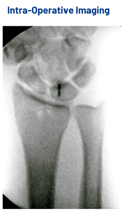
Definitive fixation
The appropriate cannulated drill was used to drill in bi-cortical fashion across the fracture site. A 2.0mm x 18mm Micro Screw was then placed across the first K-wire. The trans-articular K-wire was backed out as planned, and the screw advanced with good purchase obtained in the Bennett’s fragment and compression achieved. The trans-articular K-wire was then replaced and a second cannulated screw was advanced through a separate small stab incision and placed after drilling. As commonly seen in Bennett’s fractures, the fragment narrowed distally and required a smaller 1.5mm x 18mm Micro Screw. As this was the first time this technique was described, given the novel use of the Micro screws, it was elected to retain a single percutaneous trans-articular K-wire. However, it is now noted by Dr Larson that this is likely not necessary and since this case a variation to the technique has been adopted without the use of K-wire augmentation.
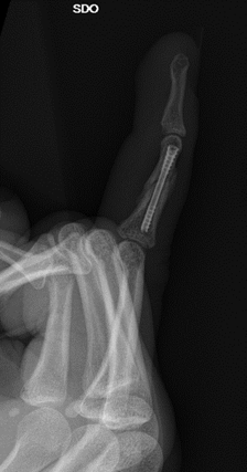
Below: Early post-operative imaging


Closure and post-operative care
The patient was placed in a thumb spica splint in theatre and as a precaution was transitioned to a cast ten days after surgery. The remaining K-wire was removed five weeks postoperatively and the patient began therapy with a removable thumb spica brace for comfort for an additional three weeks.
FOLLOW UP
At follow-up, ten weeks postoperatively, X-rays of the right thumb showed stable hardware placement and a minimally visible fracture line. The patient had excellent active range of motion and no pain or tenderness. He was allowed to advance activities to tolerance. Clinical pictures showed equal motion between the operative and contralateral thumb.
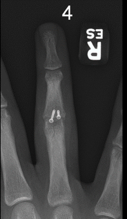
Below: Final post-operative imaging


Below: Clinical pictures illustrating motion


DISCUSSION
In this case, Dr Larson was able to achieve anatomical reduction in an unforgiving joint prone to degeneration, while addressing an injury pattern that frequently inflicts significant morbidity in the form of pain, weakness and immobilisation. Whilst this case report describes the first time the Field Orthopaedic Micro Screw was used in this way, additional precautions were taken. It is considered that the use of the Micro Screw has shown to provide a minimally invasive, rigid repair which can expedite the patient’s return to function.
The key benefits of the Micro Screw in the management of base of thumb fractures is considered to be from the small scale and cannulated features of the implant. These attributes provide the ability to take advantage of close reduction maneuvers, followed by achieving a definitive repair through a minimal incision. As the ligamentous structure around the base of the thumb is complex, and critical to biological as well as biomechanical function, reducing the operative insult is considered favorable. In addition, as the risk of sensorineural complications have been well documented and the risk is increased with the size of the incision, the minimally invasive benefits of this technique are noted.
As the thumb is a key structure of the hand and as Bennett’s fractures are one of the most common injuries, the promising outcomes seen with this new technique are considered significant. It has been well documented that fractures to the base of the thumb can be complicated to manage with significant morbidity and long-term consequences. This case highlights the Micro Screw as a highly beneficial modern solution which was able to address a number of common contributors to complications and reliably return the patient to normal function.
Product Resources
Field Orthopaedics. (2023). Micro Screw Long Surgical Technique. Brisbane, Australia: Field Orthopaedics.
FO-003275-MM Version 2 Dec 2021