MEET THE SURGEON

Dr. Libby Anderson, MD
BCom BSc MBBS FRACS FAOA
- Subspecialist Orthopaedic Hand and Wrist Surgeon
- Public and Private Practice
- Active in Research & Academic Teaching
- Special interest in trauma, nerve compression syndromes and management of carpal instability
OVERVIEW
A 35-year-old male presented to the emergency department after sustaining a crush injury to his left hand while moving heavy machinery at work. Three days later he presented to Dr Anderson for specialist review and management. Based on the clinical examination and radiographic findings, Dr Anderson performed surgery the following day. Anatomic reduction and definitive fixation were achieved by using single intramedullary 3.0 NX Nails from Field Orthopaedics for each of the proximal phalanges of the 2nd-5th fingers.
CASE INTRODUCTION
The patient is a 34-year-old right hand dominant contract labourer who presented to the emergency department (ED) after sustaining a crush injury to his left hand while moving heavy machinery at work. Radiographs confirmed significantly comminuted fractures to the proximal phalanges of the index, middle, ring, and little fingers. CT scans showed no intra-articular extension of any of the fractures.
CASE PRESENTATION
The patient presented with an extremely swollen left hand, and the range of motion (ROM) in the fingers was minimal. On examination, there were superficial grazes on the dorsal aspect of the index and little finger and volar aspect of the index finger. All wounds appeared superficial and not compound in nature. Sensation in the fingers was intact and the patient was taking oral antibiotics. On clinical examination the ring finger in particular presented with malrotation.
Below: Images upon presentation to ED

The patient’s injury was significant and the aims of treatment were to reduce swelling and promote early ROM. Additionally, employment and lifestyle factors were also considered with the patient keen to return to rugby league as a leisure activity when able. The ring finger was clearly indicated for fixation given it's malrotation. Due to these factors, the decision was made to obtain intramedullary fixation of all fractures, which would cause minimal further soft tissue trauma.
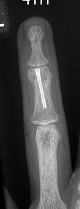
Below: Pre-Operative Imaging

PRE-OPERATIVE PLAN
Fixation of all four phalanges would provide bony stability and allow for early active ROM. The surgical plan was to fix the fractures via intramedullary nailing to avoid further soft tissue trauma. A single night stay post-surgery with intravenous antibiotics and hand therapy in the post-operative period of 6-12 weeks was recommended.
SURGICAL APPROACH
The patient presented for surgical fixation the following day, a total of four days after the initial injury. Preoperative templating was performed, and an estimated canal diameter and corresponding nail size was obtained. The procedure took place under general anaesthesia with the patient in a supine position. The wound to the volar aspect of the index finger was explored and a wash out procedure performed.
Reduction
Identical procedural steps were performed to the index, middle, ring and little fingers, in that order. The extensor mechanism was split centrally and retracted to provide visibility. Closed reduction was achieved with traction and manual pressure for all fractures. The metacarpophalangeal joints (MCPJ) were flexed and entry points identified on the dorsal third of all proximal phalanges. K-wires were passed through the intramedullary canal in an antegrade direction. Nail length was measured using the depth gauge and confirmed by holding the nail over the finger under image intensifier (II) before insertion.
Templating
To confirm the diameter, the depth gauge was aligned with the borders of the isthmus of the phalanx under fluoroscopy. When templating, there was a slight lucency surrounding the depth gauge at the isthmus to allow the nail to pass through without making contact with cortical bone. In this case, 3.0 NX Nails from Field Orthopaedics were used, ensuring subtle lucency around the threads.
Fixation
The corresponding cannulated drill bit for the 3.0 NX Nails from Field Orthopaedics was placed over the K-wire and an insertion tunnel was drilled. The drill was passed beyond the isthmus and the position of the K-wire was maintained as the drill was removed.
The standard meta drill was used (rather than the extended drill) to prepare the bone in the shape of the implant head.
After the appropriate path for the NX nail had been created, the implant was inserted in an antegrade fashion over the K-wire with careful attention paid to ensure maintenance of reduction. Correct implant placement was confirmed with fluoroscopy and rotation of the fingers carefully assessed.
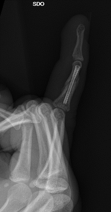
Below: Intra-Operative Imaging


Closure
5-0 prolene suture was used to repair the capsule and extensor tendons and 5-0 nylon suture used to close the wounds. Dressings with non-adhesive gauze and a plaster were applied.
Postoperative protocol
The patient was to attend an initial post-operative review with Dr Anderson’s hand therapist the following day. Following this, the patient was advised to attend follow up reviews with Dr Anderson at 2 weeks, 6 weeks and 3 months post-op. As the patient was not local a referral was made to a regional hand therapist for regular review between scheduled follow-up appointments.
Follow Up
One day review
The patient attended hand therapy the following day prior to discharge. The focus was on wound care, oedema management and early active range of motion (AROM). A hand based thermoplastic splint was fabricated for rest and the patient provided a home exercise program (HEP) to perform prior to their next review. This consisted of regular active ROM without restriction.
Two week review
The patient presented for radiological assessment with X-ray imaging confirming satisfactory healing of all four phalanges. In addition, Dr Anderson and her hand therapist performed a thorough clinical examination to assess progress. The patient presented with full sensation, moderate oedema and clean, well healed wounds. Close to full AROM was noted with the patient able to perform close to full flexion and extension of all digits. A slight lag of the little and ring fingers was noted at the proximal interphalangeal joints (PIPJ). The patient was advised to cease use of the splint for light functional activities.
Six week review
The patient presented for further radiological assessment in addition to clinical examination by both Dr Anderson and her hand therapist. X-ray imaging at this review confirmed satisfactory progressive union across all the fractures. Clinically, the patient presented with excellent AROM. A slight lag at the PIPJ of the ring and little fingers remained evident however improved during the hand therapy session. Grip strength was recorded at 42 kilograms. Some atrophy of the patients intrinsic and first dorsal interossei muscles with weak abduction and adduction of the fingers was noted. This was likely secondary to the patients crush injury. Ongoing hand therapy was to concentrate on intrinsic strengthening. The patient returned to suitable duties at this stage, working within the limits of their pain in a manual job.
Three month review
The patient presented with excellent range of motion and strength and was cleared for full duties at this review.

Discussion
Dr Anderson was able to treat a significant injury for a young, active patient and achieve full return to function within three months using the NX Nail from Field Orthopaedics. Definitive fixation through a percutaneous approach minimised further soft tissue disruption and provided the patient an expedited return to full function and a complete recovery.
References
1. Field Orthopaedics. (2023). NX Nail Product Brochure. Brisbane, Australia: Field Orthopaedics.
2. Field Orthopaedics. (2024). NX Nail Phalanx Long Surgical Technique. Brisbane, Australia: Field Orthopaedics.
FO-009936-MM Version 1 Oct 2024