MEET THE SURGEON

Dr Andrew Mayo, MD
BSc(Med) MBBS(Hons) MEng FRACS FAOrthA
Subspecialist Upper Limb Surgeon Public and Private Practice Active in Teaching
Special interest in hand, wrist, elbow, and shoulder trauma, degenerative conditions and instability
DISCLAIMER
The opinions expressed by Dr. Mayo are those of Dr. Mayo and not necessarily those of Field Orthopaedics. Individual experiences may vary.
The procedure described in the report may differ from the manufacturer’s surgical technique. Surgeons are advised to review the product specific surgical technique prior to performing any surgery.
A surgeon must always rely on their own professional clinical judgement when deciding whether to use a particular product when treating a particular patient. A surgeon must always refer to the instructions for use, product label and surgical technique before using any Field Orthopaedics product. Product may not be available in all markets. Please contact your Field Orthopaedics representative if you have any questions about availability of Field Orthopaedics products in your area.
OVERVIEW
A 51-year-old right hand dominant office worker presented to clinic after sustaining an injury to her left hand after a collision with a tree while riding a time trial bike. Radiographs confirmed displaced shaft fractures of the 2nd, 3rd, 4th and 5th metacarpals. The skin was not breached and the decision was made to perform percutaneous reduction and internal fixation using NX Nails from Field Orthopaedics.
CASE INTRODUCTION
The patient was a 51-year-old right hand dominant office worker who presented to clinic for evaluation of a closed injury to her left hand. The injury was sustained after a collision with a tree while riding a time trial bike and radiographs confirmed displaced shaft fractures of the 2nd, 3rd, 4th and 5th metacarpals.
CASE PRESENTATION
On examination, there was generalised swelling over the dorsum of the hand and bruising in the palm. Tenderness to palpation was noted over metacarpals 2-5 and flexion was limited due to swelling. Rotation was difficult to assess due to the fact that all four metacarpals were fractured but there was some discrepancy in rotational alignment noted between digits.
Below: Pre-Operative Imaging
Imaging demonstrating displaced fractures of metacarpals 2-5

Below: Pre-Operative Imaging
Imaging demonstrating displaced fractures of metacarpals 2-5

PRE-OPERATIVE PLAN
The patient was otherwise healthy and desired to return to activity as soon as possible, therefore percutaneous reduction and internal fixation was recommended. Due to the nature and extent of the injury, the patient was advised the procedure may require open reduction to achieve adequate fixation.
SURGICAL APPROACH
The patient presented for surgical fixation five days after the injury. Preoperative templating was performed and an estimate of canal diameter and corresponding nail size was obtained. The procedure took place under general anaesthetic with the patient in supine position.
Reduction
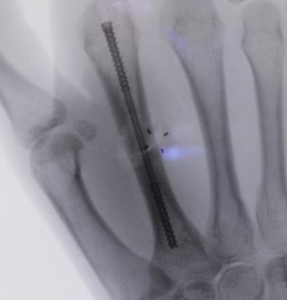
Using the retrograde technique the metacarpophalangeal joints (MCPJ’s) were flexed to 90 degrees and the percutaneous entry points identified within the dorsal third of each metacarpal head. Under fluoroscopy, distraction and indirect pressure was used to anatomically reduce each of the fractures and working radial to ulna, K-wires were inserted longitudinally into the medullary canal of each metacarpal. K-wire insertion started at the centre of the metacarpal head, in the coronal plane and at the dorsal 1/3 of the metacarpal head in the sagittal plane. K-wires were progressed across the fracture site and into the base of the metacarpals to the depth of their final position. This ensured correct length measurement for the NX Nails. Acceptable closed reduction was achieved for all metacarpals.
Templating
To confirm the diameter, the depth gauge was aligned with the borders of the isthmus of the metacarpals under fluoroscopy. When templating, there was a slight lucency surrounding the depth gauge at the isthmus to allow the nail to easily pass through without making contact with cortical bone.
In this case, a 3.5mm NX Nail was used for the 2nd and 3rd while a 3.0 mm NX Nail was used for the 4th and 5th metacarpals, ensuring subtle lucency around the threads.
Fixation
The corresponding cannulated drill bits for the 3.0mm and 3.5mm Field Orthopaedic NX Nails were placed over the K-wires and insertion tunnels were drilled. The drill was passed beyond the isthmus and the position of the K-wire was maintained as the drill was removed. The standard metaphyseal drill was then used to help prepare the bone in the shape of the implant head.
After the appropriate path for the NX Nails had been created, the implants were inserted in a retrograde fashion over the K-wires with careful attention paid to the maintenance of the anatomic reduction of the fracture site. Correct implant placement was confirmed with fluoroscopy and rotation of each finger carefully scrutinised and noted to be anatomic relative to the digit cascade of the patients contralateral unaffected hand. Normal tenodesis of the digital flexors and extensors of all operative fingers was noted.

Closure
A single 5-0 nylon suture was used to close the entry wound for each metacarpal. Dressings with non-adhesive gauze and a volar back slab were applied.
Postoperative protocol
The patient was referred to hand therapy for a protective mallet splint and encouraged to start full distal interphalangeal joint (DIPJ) range of motion immediately.
Follow Up
The patient was reviewed at two weeks and three months post operatively.
Two week review
At the two week post operative appointment the small wounds from the percutaneous entry points were well healed. The patient had attended hand therapy following the procedure which focussed on active mobilisation and oedema control. Splint wear was protective only and the patient was able to make a full composite fist.

Three month review
At the standard three month post operative review, the patient reported no pain, had weaned from splint use and had returned to normal daily activities and work tasks. The patient was deemed to have completed a full clinical recovery.


References
1. Field Orthopaedics. (2022). NX Nail Product Brochure. Brisbane, Australia: Field Orthopaedics.
2. Field Orthopaedics. (2023). NX Nail Long Surgical Technique. Brisbane, Australia: Field Orthopaedics.
FO-008563-MM Version 1 July 2024