MEET THE SURGEON

Dr Sagar Shah, MD
Orthopedic Surgery Residency Louisiana State University New Orleans, LA
Hand, Upper Extremity, and Microvascular Surgery Fellowship, University of Colorado.
Doctor of Medicine, Virginia Commonwealth University
OVERVIEW
The patient was a 22-year-old female who is 9 weeks pregnant, right-hand dominant, and working as a housekeeper. The patient sustained multiple right upper extremity fractures in an Motor Vehicle Crash (MVC) in May 2023. The patient sustained a both-bone forearm fracture and underwent Open Reduction and Internal Fixation (ORIF) by the orthopedic trauma team. The patient also sustained right fourth and fifth metacarpal fractures. She was evaluated in clinic by Dr. Shah where she was found to have a clinically significant extension lag of the 4th digit. Dr. Shah and the patient discussed operative versus nonoperative treatment and went over the risks of both. The patient elected for operative treatment.
CASE INTRODUCTION
The patient was a 22-year-old female who was 9 weeks pregnant, right-hand dominant, and working as a housekeeper. The patient sustained multiple right upper extremity fractures in an MVC in May 2023.
The patient sustained a both-bone forearm fracture and underwent ORIF by the orthopedic trauma team. She also sustained right fourth and fifth metacarpal fractures. She was evaluated in clinic by Dr. Shah where she was found to have a clinically significant extension lag of the fourth digit. Dr. Shah and the patient discussed operative versus nonoperative treatment and went over the risks of both. The patient elected for operative treatment.
CASE PRESENTATION
On examination, swelling was observed over the dorsal radial hand. There was bruising in the palm and there was tenderness to palpation over the fourth metacarpal shaft and fifth metacarpal base. The fourth metacarpal head was prominent and palpable in the palm. There was an extension lag of the ring finger. Flexion was limited due to swelling in the hand. There was no rotational deformity. Prior to this injury, she had no pain, disability, extension lag, or weakness in this hand.
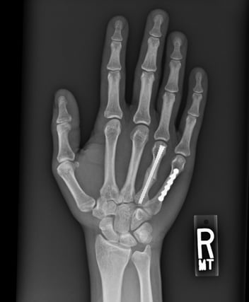
Below: Pre-Operative Imaging

Radiographically, there was a transverse fracture of the fourth metacarpal shaft with a comminuted fracture of the fifth metacarpal base. The fifth carpometacarpal joint appeared to be congruent. The second metacarpal was shortened (approximately 5mm) and flexed (approximately 45 degrees).
PRE-OPERATIVE PLAN
Surgical and nonsurgical treatment options were discussed with the patient. Given the shortening and flexion deformity of the metacarpal, closed treatment without manipulation was not an acceptable option. Closed reduction and casting were presented as an option with the risk of loss of reduction and malunion. The patient was made aware that a malunion of this severity may cause permanent dysfunction in the hand. The patient was a young healthy female who desired to return to activity as soon as possible.
For these reasons, the decision was made to proceed surgically with anatomic reduction of the fracture and definitive fixation with an NX Nail from Field Orthopedics to get her moving again and reduce the risk of long-term dysfunction of the ring finger.
SURGICAL APPROACH
The patient presented for surgical fixation seven days after the injury.
Preoperative templating was performed. In this case a CT scan was obtained to confirm that the fifth carpometacarpal joint was congruent. As the scan provided relevant information and had already been performed, it was also then used to template the NX Nail measurement. The canal measured 2.0mm so a 2.0mm nail diameter was selected.

Reduction
Appropriate reduction was obtained.
Templating
A 2.0mm NX Depth Gauge was used to verify the diameter of the implant required.
Ensuring “BONE DIAMETER CAN BE MEASURED HERE” was facing up, the edges of the depth gauge were aligned with the borders of the isthmus of the metacarpal while under fluoroscopy.
Slight lucency surrounded the depth gauge at the isthmus which ensured the diameter was not too large.

A second technique to template the NX Nail size was also performed by using the NX Nail itself. A nail of the templated diameter, in this case a 2.0mm nail, was selected and was placed over the metacarpal and diameter visualized under fluoroscopy. The distal threads were placed at the isthmus. There was a subtle lucency around the distal threads at the isthmus confirming correct size selection.

Fixation
A K-wire was placed through the open traumatic wound in the distal metacarpal at the level of the metacarpal head. The starting point was the centre of the metacarpal head on the AnteriorPosterior (AP) view and at the dorsal ⅓ of the metacarpal head in the lateral view.
Intraoperative fluoroscopy was utilized to advance the K-wire in a retrograde fashion into the medullary canal, across the fracture site, into the base of the second metacarpal and confirm anatomic reduction and desired K-wire placement.


The corresponding 1.5mm cannulated drill for the 2.0mm Field Orthopedics NX Nail was used to drill over the K-wire via the open incision. Since the fourth metacarpal had an especially narrow canal, the cannulated drill (1.75mm) for the next size up NX Nail (2.5mm) was also passed across the isthmus to ream the isthmus and allow for easier nail passage.

In the tray, the standard metaphyseal drill was located and used to prepare the bone in the shape of the implant head and alleviate the forces generated by it. The metaphyseal drill was passed over the K-wire and used to over drill the insertion tunnel. Again, care was taken to maintain the position of the K-wire as the drill was removed.


After the appropriate path for the nail had been created, a 2.0mm x 40mm NX Nail was inserted in a retrograde fashion over the K-wire with careful attention paid to maintenance of the anatomic reduction of the fracture site. Correct implant placement was confirmed with fluoroscopy and rotation of the finger carefully scrutinized and noted to be anatomic relative to the cascade of the adjacent digits as well as her contralateral unaffected hand. Normal tenodesis of the digital flexors and extensors of the ring finger was noted.

In this case, the fifth metacarpal was fixed to the fourth metacarpal with 0.045 K wires.

Closure
The wounds were irrigated and closed with 4-0 nylon suture in a horizontal mattress fashion before being dressed with sterile Xeroform, 4x4 gauze, cast padding and a well-padded forearm based volar wrist splint.
Postoperative Protocol
Patient was encouraged to work on active range of motion in the splint immediately.

FOLLOW UP
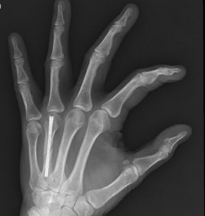
Eight weeks postoperative
Radiographically, the fourth metacarpal fracture had healed in anatomic alignment.
Clinically, the patient had regained full range of motion of her ring finger without any malrotation or extensor lag. She returned to all activities that she enjoyed doing including weightlifting.

References
1. Field Orthopaedics. (2022). NX Nail Product Brochure. Brisbane, Australia: Field Orthopaedics.
2. Field Orthopaedics. (2023). NX Nail Long Surgical Technique. Brisbane, Australia: Field Orthopaedics.
FO-008563-MM Version 1 July 2024