MEET THE SURGEON

Dr Raymond K. Wurapa, MD
Orthopedic Surgeon, Orthopedic ONE,
Columbus Ohio.
Specialist in hand, wrist and upper extremity surgery
Fellow, Philadelphia Hand To Shoulder Center,
Thomas Jefferson University (2000)
OVERVIEW
A 37-year-old male sustained a crush injury to his non-dominant hand at work. He was initially evaluated at a local emergency department before presenting to Dr Wurapa three days later. Upon evaluation, he had a dorsal laceration and a fifth metacarpal fracture of the right hand which involved the distal diaphysis with comminution.
After discussing operative and non- operative treatment options for the fracture, the patient elected to proceed with open reduction internal fixation to minimize limitations during his recovery and return to work. Dr Wurapa achieved definitive fixation with a 4.0mm diameter, 45mm length NX Nail following debridement and repair of the dorsal wound. By four weeks post-operatively the patient reported no pain, had weaned splint use, and returned to normal daily activities and work tasks with full digital motion. At his final appointment he was deemed to have completed a full clinical recovery with solid consolidation at the fracture site. In this case, the NX Nail offered Dr Wurapa a solution for treating a complex fracture while maintaining length, restoring version, and controlling angulation1.
CASE INTRODUCTION
A 37-year-old male sustained a crush injury to his non-dominant hand at work from falling equipment. He was initially evaluated at a local emergency department and noted to have a dorsal hand laceration and a metacarpal fracture. The wound was irrigated and loosely closed before a short arm splint was applied. The patient was referred to Dr Wurapa for consultation and definitive management.
CASE PRESENTATION
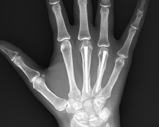
The patient presented to Dr Wurapa three days after being evaluated at the local emergency department. Upon evaluation, he had pain and swelling at the point of injury, and the dorsal hand wound was determined to require additional debridement. Radiographs revealed a fifth metacarpal fracture of the right hand which involved the distal diaphysis with comminution.
Below: Pre-Operative Imaging


PRE-OPERATIVE PLAN
After discussing operative and non-operative treatment options for the metacarpal fracture, the patient elected to proceed with definitive fixation to minimize limitations during his recovery and return to work as soon as possible. Dr Wurapa planned to use the NX Nail from Field Orthopaedics as he believes the anatomic design to be ideal for this distal fracture pattern due to the bone capture it achieves through the head and neck region. The non-compressive design also avoids unnecessary compression and shortening in the presence of comminution. Additionally, the dorsal hand laceration would be debrided.
After discussing operative and non-operative treatment options for the metacarpal fracture, the patient elected to proceed with definitive fixation to minimize limitations during his recovery and return to work as soon as possible. Dr Wurapa planned to use the NX Nail from Field Orthopaedics as he believes the anatomic design to be ideal for this distal fracture pattern due to the bone capture it achieves through the head and neck region. The non-compressive design also avoids unnecessary compression and shortening in the presence of comminution. Additionally, the dorsal hand laceration would be debrided.
OPERATIVE APPROACH
Five days after the initial consultation, the patient underwent successful reduction and fixation of the fifth metacarpal fracture following debridement and repair of the dorsal hand laceration.
Under a regional block anaesthesia with light sedation, the arm was prepped and draped per routine. The extremity was exsanguinated, and upper arm tourniquet raised to 200mm Hg.
Under mini C-arm fluoroscopy the NX Nail depth gauge was used to identify the appropriate nail diameter. A guidewire was placed retrograde under power and advanced through an entry point at the dorsal third midline position of the metacarpal head. The guidewire was advanced to the proposed depth of the nail within the fifth metacarpal.
A 5mm longitudinal skin incision was made above and below the guidewire. This allowed introduction of the depth gauge to determine the appropriate nail length. The guidewire was then advanced under power to avoid inadvertent extraction during the remainder of the case. The cannulated drill was passed over the guidewire with required depth confirmed under fluoroscopy. A 4.0mm diameter, 45mm length NX Nail was then advanced by hand over the guidewire and seated into place. Care was taken with final seating of the implant to verify clinical alignment of fifth digit and reduction at the fracture site.
The guidewire was extracted, final images obtained, and the wound closed with a 5-0 prolene suture. A light soft dressing was placed on the hand with instructions to begin immediate gentle mobilization.


FOLLOW UP
Due to the hand laceration, the patient was seen one week post-operatively, at which time all wounds were healing well. He continued active mobilization of his hand and returned to work using an ulnar gutter splint for heavy activity only. A brief course of therapy was initiated to create a home exercise program focused on oedema control and maximal digital mobilization. By four weeks post-operatively the patient reported no pain, had weaned all splint use, and had returned to normal daily activities and work tasks. At his final follow up appointment eight weeks after the procedure, he was deemed to have completed a full clinical recovery with solid consolidation at the fracture site.


DISCUSSION
With an anatomically contoured compaction taper head which has been designed from statistical shape models, the NX Nail maximises bone engagement and distal fragment control to provide stability in the management of comminution1. Additionally, the non-compressive design maintains length while avoiding any unnecessary compression at the fracture site. Manufactured from Titanium alloy, the NX Nail can withstand normal loads and facilitate a rapid return to normal activity by acting as a physiological splint1. The combination of a high precision leading tip, smooth shaft and anatomically contoured head facilitate the stabilization of key bone structures in an anatomic position to restore native biomechanics1. In this case, the NX Nail offered Dr Wurapa a solution for treating a complex fracture while maintaining length, restoring version, and controlling angulation1, enabling his young patient to quickly return to normal daily activities and work tasks without restriction.
References
1. Field Orthopaedics. (2022). NX Nail Design Rationale. Brisbane, Australia: Field Orthopaedics.
FO-005737-MM Version 2 Nov 2023 6