
MEET THE SURGEON

Dr Sagar Shah, MD
Orthopedic Surgery Residency, Louisiana State University, New Orleans, LA.
Hand, Upper Extremity, and Microvascular Surgery Fellowship, University of Colorado.
Doctor of Medicine, Virginia Commonwealth University
DISCLAIMER
The opinions expressed by Dr. Shah are those of Dr. Shah and not necessarily those of Field Orthopaedics. Individual experiences may vary.
The procedure described in the report may differ from the manufacturer’s surgical technique. Surgeons are advised to review the product specific surgical technique prior to performing any surgery.
A surgeon must always rely on their own professional clinical judgement when deciding whether to use a particular product when treating a particular patient. A surgeon must always refer to the instructions for use, product label and surgical technique before using any Field Orthopaedics product. Product may not be available in all markets. Please contact your Field Orthopaedics representative if you have any questions about availability of Field Orthopaedics products in your area.
OVERVIEW
A 22-year-old male presented to Dr Shah after sustaining an injury to his right hand after he dropped a dresser while moving house. Radiographs confirmed there was a short oblique fracture of the second metacarpal shaft.
With a desire to return to his active lifestyle, the decision was made to proceed surgically with anatomic reduction of the fracture and definitive fixation with an NX Nail from Field Orthopaedics. The use of the NX Nail provides the patient with stability, allows rapid return to function, and reduces the risk of deformity1. The patient was encouraged to work on active range of motion in the splint immediately after surgery.
CASE INTRODUCTION
The patient is a 22-year-old right-hand dominant male with a history of sickle cell disease. He presented to Dr Shah for evaluation of a closed fracture of the second metacarpal shaft after he dropped a dresser on his hand while moving house.
CASE PRESENTATION
On examination, there was swelling over the dorsal radial hand. There was bruising in the palm. There was tenderness to palpation over the second metacarpal shaft. The second metacarpal head was prominent and palpable in the palm. There was an extension lag of the index finger. Flexion was limited due to swelling in the hand. There was no rotational deformity. Prior to this injury, he had no pain, disability, extension lag, or weakness in his hand.
Below: Pre-Operative Imaging
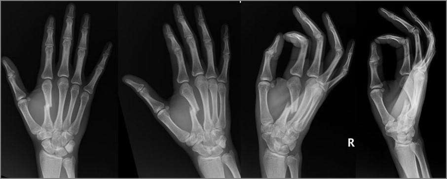
Radiographically, there was a short oblique fracture of the second metacarpal shaft. The second metacarpal was shortened (approximately 8mm) and flexed (approximately 48 degrees).
PRE-OPERATIVE PLAN
Both surgical and non-surgical treatment options were discussed with the patient. Given the shortening and flexion deformity of the metacarpal, closed treatment without manipulation was not an acceptable option. Closed reduction and casting were presented as an option with the risk of loss of reduction and malunion. The patient was made aware that a malunion of this severity may cause permanent dysfunction in the hand.
The second metacarpal can have a surprisingly narrow isthmus in a young, fit male. The patient was otherwise healthy and desired to return to activity as soon as possible.
For these reasons, the decision was made to proceed surgically with anatomic reduction of the fracture and definitive fixation with an NX Nail from Field Orthopaedics to get him moving again and reduce the risk of long-term dysfunction of the index finger. As the NX Nail has been designed with an anatomically inspired head, increased distal engagement and a non-compressive design (based on the AO principles of fracture management), it was considered to provide stability, allow rapid return to function, and reduce the risk of deformity1.
SURGICAL APPROACH
The patient presented for surgical fixation seven days after the injury. Preoperative templating was performed. Although a radiographic marker ball was not used to calibrate an exact measurement, an estimate of canal diameter was obtained. In this case, canal diameter was measured to be 2.5mm.


Reduction
An attempt at a closed reduction with longitudinal traction and manipulation of the fracture was unsuccessful. A K-wire was used to percutaneously reduce the fracture, but this was also unsuccessful. A 2 cm incision was made over the fracture site. Dissection was carried through subcutaneous tissue with care to protect branches of the radial sensory nerve. The index extensor tendons were protected and retracted in an ulnar direction. A freer was used to shoehorn the fracture into a reduced position.
Templating
A Field Orthopaedics 2.5mm Depth Gauge was used to determine the diameter of the implant required.
To confirm the diameter, the depth gauge was aligned with the borders of the isthmus of the metacarpal under fluoroscopy. When templating, there was a slight lucency surrounding the depth gauge at the isthmus to allow the nail to easily pass through. Attempting to pass an oversized nail can cause propagation of the fracture, iatrogenic fracture, or fracture of the screw.


In this case, a 2.5mm nail was selected and placed over the second metacarpal. The distal threads were placed at the isthmus, ensuring there was a subtle lucency around the threads.


Fixture
A guidewire was placed percutaneously in the distal metacarpal at the level of the metacarpal head. The wire started at the center in the metacarpal head on the AP view and at the dorsal ⅓ of the metacarpal head in the lateral view. Intraoperative fluoroscopy was utilised to advance the guidewire in a retrograde fashion into the medullary canal, across the fracture site.
The Field Orthopaedics depth gauge was used to determine implant length. The K-wire was then advanced into the base of the second metacarpal and confirm anatomic reduction and desired guidewire placement. A 1cm incision was made over the guidewire at the distal aspect of the metacarpal.


The corresponding drill for the 2.5mm Field Orthopaedics NX Nail was used to drill over the guidewire via the open incision.
There are two metaphyseal drills, the standard and extended, to help prepare the bone in the shape of the implant head and alleviate any forces generated by it. The standard meta drill was used in this case, identified by the single colour band on the shaft and the absence of a laser-marking band on the head. The bone was prepared by passing the meta drill over the K-wire and over-drilling the insertion tunnel. The position of the K-wire was maintained as the drill was removed.


After the appropriate path for the nail had been created, a 2.5mm x 60mm NX Nail was inserted in a retrograde fashion over the guidewire with careful attention paid to the maintenance of the anatomic reduction of the fracture site. Correct implant placement was confirmed with fluoroscopy and rotation of the finger carefully scrutinised and noted to be anatomic relative to the cascade of the adjacent digits as well as the patient’s contralateral unaffected hand. Normal tenodesis of the digital flexors and extensors of the index finger were noted.


Closure
The wounds were irrigated and closed with 4-0 nylon suture in a horizontal mattress fashion before being dressed with sterile Xeroform, 4x4 gauze, cast padding and a well-padded forearm based volar wrist splint.
Postoperative protocol
The patient was encouraged to work on active range of motion in the splint immediately.
References
1. Field Orthopaedics. (2022). NX Nail Product Brochure. Brisbane, Australia: Field Orthopaedics.
2. Field Orthopaedics. (2023). NX Nail Long Surgical Technique. Brisbane, Australia: Field Orthopaedics.
FO-006364-MM Version 2 Nov 2023


