MEET THE SURGEON

Dr Jordan Grier, MD
Orthopedic Surgeon in Cleveland, Ohio
Specialising in upper extremity (hand, wrist, elbow and shoulder).
Assistant Professor of Orthopaedic Surgery,
Case Western Reserve University School of Medicine.
Medical Degree and Residency Training,
Duke University Medical Centre.
Subspecialty Training,
OrthoCarolina Hand Centre.
OVERVIEW
A 25-year-old right-hand-dominant male presented with a high energy injury to the right wrist after a motorcycle accident. X-rays showed fractures of the distal radius, distal ulna, scaphoid and hook of hamate.
The patient was treated with a combination of the Field Orthopaedics 4.0mm NX Extremity Nail to provide fixation to the distal ulna, a dorsal bridge plate to temporarily stabilize and maintain the length of the distal radius and parallel headless compression screws with bone graft for the scaphoid waist fracture.
CASE INTRODUCTION
A 25-year-old male industrial worker was admitted to inpatient care after suffering numerous injuries including pulmonary contusions, facial and multiple wrist fractures, following a motorcycle accident. The patient was seen and operatively managed by hand surgery five days post injury. Imaging of the right wrist showed a three column, extensively comminuted and impacted distal radius fracture; a displaced distal ulna neck fracture; a displaced scaphoid waist fracture and a minimally displaced fracture to the hook of hamate.
CASE PRESENTATION
The patient was initially evaluated at UH Cleveland Medical Centre and admitted due to the extensive injuries. On examination in the ward by Orthopaedics, the patient remained in a well-padded thumb spica sugar-tong splint, applied to the right wrist during admission by the orthopaedic surgery consultant. There was mild swelling, and the wrist was diffusely tender to palpation. The hand was well perfused and neurovascularly intact.
Below: Pre-Operative imaging


PRE-OPERATIVE PLAN
Due to the extensive nature of the right wrist injuries and the need for stabilization, Dr Grier admitted the patient for Open Reduction and Internal Fixation (ORIF) of the right distal radius and scaphoid fractures, with the intent to decide intraoperatively on the requirement to fix the distal ulna.
As with other fractures the intent for treating high energy wrist injuries involves restoring length and alignment, whilst attempting to obtain congruency of the articular surface, particularly of the radiocarpal and distal radioulnar joints. Unfortunately, unlike simpler fracture patterns, high energy injuries are often associated with extensive articular surface comminution and extension into the radial diaphysis, as was the situation in this case.
Multiple treatment options are available for the management of high-energy distal radius fractures including locked volar plating constructs, dorsal plating constructs, external fixation, and temporary dorsal spanning fixation. In this case given the extent of articular comminution present, Dr Grier elected to manage the distal radius injuries with the use of a temporary dorsal spanning plate to maintain length and alignment through bridging whilst healing occurs. This fixation methods allows for excellent reduction of the comminuted fracture fragments without the need for extensive handling or stripping of the soft tissues present about the fracture.
For the scaphoid, due to the displaced nature of the waist fracture, it was considered that the 70–80% of inter-osseous blood supply was impacted and the viability of the proximal scaphoid would be compromised without repair. As the scaphoid unites through primary bone healing, as seen in other intra-capsular fractures, Dr Grier opted for rigid fixation with two parallel headless compression screws inserted anterograde for convenience due to the other exposures required in the vicinity of the dorsal wrist.
The hook of hamate fracture was assessed on both plain radiographs and CT scan and noted to be nondisplaced. Dr Grier discussed with the patient the possibility that this fracture may fail to unite, and if so, could be excised at a later date if symptomatic.
Lastly, whilst Dr Grier initially elected to make a decision regarding ulna fixation during the operation, the preoperative plan considered that if this was to occur it would be by using the 4.0mm Field Orthopaedics NX Nail. The considerations for using this implant related to the impressive strength of the construct, the simplicity of the procedure and the compatibility of the titanium nail with the other titanium implants chosen to fix the remainder of the fractures.
Distal ulna fixation via the K-wire guided, retrograde insertion of an NX Nail is considered to be an extremely fast and simple way to achieve a strong repair. Due to the intramedullary technique, the majority of the soft tissue is preserved and the non-compressive design is very effective at restoring the length of the ulna, hence stabilising the wrist and reconstructing the Distal Radioulnar Joint (DRUJ). As most high energy wrist injuries often have numerous fractures and require multiple implants, there is a risk of dissimilar metal reactions. Whilst it might not be commonly considered an additional noteworthy benefit, titanium is observed to be both highly biocompatible and compatible with other implants. In addition to the hypoallergenic and osteogenic properties of titanium, it is often used in many implants and hence allows a greater variety of implant choices without the concern of galvanic corrosion.
SURGICAL APPROACH
Due to the extensive nature of injuries sustained by this patient, the single operation was managed in three stages. The order of approach involved first fixing the scaphoid whilst able to flex the wrist. Then bridging the radius to restore length and alignment and allow the initial management to be radiographically assessed. Finally, it was decided to operatively repair the ulna in order to reconstruct the DRUJ.
Below: Intra-operative imaging


Stage One: Scaphoid
To commence the operation, a dorsal incision was made over the wrist between the third and fourth extensor compartments. Blunt dissection was carried through the subcutaneous tissues to the level of the extensor retinaculum with great care taken to preserve the traversing branches of the Superficial Branch of the Radial Nerve (SBRN). The extensor retinaculum was visualized and incised overlaying the Extensor Pollicis Longus (EPL) tendon and the tendon was translocated and protected with self-retaining retractors. The dorsal wrist capsule was identified and incised in line with the fibers of the dorsal radiocarpal ligament.
The dorsal surfaces of the proximal pole of the scaphoid, lunate and scapholunate interosseous ligament were identified. The axis of the scaphoid was initially estimated by identifying the axis of the scaphoid-lunate plane. A K-wire was then placed along the long-axis of the scaphoid, and desired position confirmed with orthogonal fluoroscopic views. A second K-wire was placed in a parallel fashion.
Once Dr Grier was satisfied with the K-wire position, screw length was measured and the K-wire over drilled in preparation for an 18mm cannulated screw which was placed in standard fashion. It was noted that correct screw length is always 4–5 mm shorter than measured to avoid protrusion of the screw. Screw alignment and position was confirmed by fluoroscopy at multiple angles before closure (and in this case before bridge plating) to make sure there was no protrusion, and that passive motion of the wrist could be achieved without hindrance.
Once the first screw was inserted, cancellous autograph was harvested from the radial metaphysis and placed via a second screw hole into the fracture void distally in the scaphoid. A second 18mm cannulated screw was placed in standard fashion over a guidewire and placed through the graft. Desired screw positioning was confirmed again via orthogonal fluoroscopic views.
Stage Two: Distal Radius
With the assistance of fluoroscopy, the displaced fragments of the distal radius were reduced with a combination of longitudinal traction and palmar translation to restore radial length, inclination and sagittal tilt.
In preparation for plating, the dorsal bridging plate was superimposed over the radial diaphysis to the diaphysis of the third metacarpal and verified with a C-arm. The skin at the level of the proximal and distal screw holes was marked. A 3cm incision was made over the dorsum of the third metacarpal base and sharp dissection carried through to the level of the Extensor Digitorum Communis (EDC) to the long finger. Care was taken to preserve crossing branches of the SBRN. The long finger extensor tendon was retracted ulnarly and the dorsal cortex of the third metacarpal was exposed.
The prior dorsal wrist incision from scaphoid fixation was extended over the dorsum of the distal radius just ulna to Lister’s tubercle. Sharp dissection was carried through skin and subcutaneous tissues with the crossing branches of the SBRN preserved. The third dorsal compartment was identified, and the EPL tendon was transposed from its tendon sheath. The positioning under the sheath is considered imperative and is well cited in literature. It is considered that this placement helps avoid impingement of the extensor tendons on the plate and that failure to do so can be associated with both post-operative irritation and on occasion rupture.
With the EPL tendon protected, the fourth extensor compartment was lifted from radial to ulnar in a subperiosteal fashion with care taken not to disrupt the Posterior Interosseous Nerve (PIN) or Posterior Interosseous Artery (PIA). The fracture site was identified and debrided to facilitate anatomic reduction.
A third 3cm incision was made just proximal to the palpable first dorsal compartment muscle bellies. Sharp dissection was carried to the level of the second dorsal compartment fascia taking care to protect the Lateral Antebrachial Cutaneous Nerve (LABCN) and SBRN. The Extensor Carpi Radialis Longus (ECRL) - Extensor Carpi Radialis Brevis (ECRB) interval was developed to allow access to the radial shaft. A periosteal elevator was passed deep to the dorsal musculature from proximal to distal to ensure adequate space for the plate passage. An appropriately sized dorsal spanning plate was selected and fluoroscopy used to ensure the desired placement before screws were implanted to secure the plate in place.
Stage Three: Distal Ulna
During the procedure, the decision was made to rigidly fix the ulna fracture with the NX Nail from Field Orthopaedics due to the simplicity afforded by the implant system and the expected benefits that rigid fixation would have on the DRUJ.
A guidewire was inserted into the corresponding start point in the distal ulna head. Placement of this wire is most efficiently performed with the forearm in a fully pronated position on the operating table. Pronation of the forearm allows for subtle palmar migration of the distal radius, allowing the ulna head to rest in a more prominent position about the dorsal wrist. The percutaneous entry portal is best identified at the level of the dorsal body of the triquetrum, passing proximally in a colinear fashion with the ulna diaphysis. The correct starting point for the K-wire is achieved via placement just radial to the ulnar styloid at the level of the ulnar fovea. Visualization of the guidewire within this sulcus of the distal ulna is similar to the radiographic placement of the starting guidewire within the piriformis fossa for femoral nailing, a procedure commonly performed in orthopaedic trauma practice. Once satisfactory alignment was confirmed on orthogonal fluoroscopic views, the guidewire was advanced into the ulna diaphysis. Implant length was measured using the corresponding Field Orthopaedics measuring guide and a 4.0mm x 50mm NX Nail selected and inserted in standard fashion after drilling over the guidewire. Inserting the NX Nail reduced and fixed the fracture as well as restored the alignment of the DRUJ. The repair and placement of the hardware was confirmed on orthogonal fluoroscopic views and was shown to be satisfactory.
Below: Intra-operative imaging showing fixation


Closure
Wounds were irrigated with saline and closed in layers with nylon sutures. A well-padded volar resting splint was applied with the metacarpophalangeal joints free for uninhibited digital flexion immediately postoperatively.
SURGICAL APPROACH
The patient had mild pain in the wrist for the first three to four days, as would be expected with the extensive nature of the injuries and the operation. However, by the time of the two week review the patient reported all pain resolving and no need for pain medications. Examination of the right wrist and forearm demonstrated well-healing incisions and composite digital flexion less than 1 cm from the distal palmar crease for all digits. Anterior Interosseous Nerve (AIN), PIN and ulna motor function remained intact.
Radiographs of the right wrist demonstrated preserved post-operative alignment and hardware in place without evidence of loosening or failure. Arm and finger range of motion and hand use were encouraged with the caution to avoid heavy use, including gripping, squeezing and direct pressure on the wound. The patient was referred to hand therapy for initiation of active motion rehabilitation. Three months post-operatively the patient had clinical and radiographic union of the fracture which indicated that the dorsal spanning plate could be removed. Despite a positive COVID diagnosis delaying this removal by one month, the patient had an excellent outcome with no pain or functional impairment.
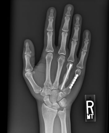
Below: Immediate Post-operative Imaging


Below: Two week post-operative imaging


Below: Four months post-operative imaging following plate removal


DISCUSSION
High energy wrist injuries create extensive and complex fracture patterns to the hand and wrist with injuries often involving fractures to the distal radius and ulna. However, depending on the energy of the injury and the position of the wrist at the time, these injuries can also include fractures to the carpus, as well as radiocarpal, perilunate and other intracarpal dislocations.
The impact of these injuries is significant. Not only is the trauma extensive with other concomitant pathologies, the patient is often young with a high functional demand. Weber and Szabo1 noted that highly comminuted distal wrist fractures such as these are unfortunately associated with a very high complication rate (52%-63%) when treated by traditional techniques. These issues have been considered to arise from the traditional use of external fixators combined with the prolonged duration required to achieve union.
Whilst large scale evidence is still not available to suggest the most appropriate way to manage these injuries, it does appear from the emerging data and short-term results of this case report and others similar, that implicitly there is merit in these modern techniques. In this report, it is demonstrated by Dr Grier how a combination of modern implants can be used under X-ray guidance and through minimally invasive incisions to effectively address what has previously been considered a very complex and difficult condition to manage.
The inclusion of the NX Nail to manage the distal ulna fracture has been shown to be an excellent addition to this management approach. To be able to so quickly and effectively restore the length and alignment of the distal ulna and concentricity of the DRUJ, has notable benefits in all high energy wrist injuries and has now been adopted as a key component in this treatment approach for future cases.
References
1. Treatment of high energy distal radius fractures using bridging plate. Available from:
https://www.researchgate.net/publication/335600512_Treatment_of_high_energy_distal_radius_fractures_using_bridging_plate [accessed Jan 19 2022].
FO-003268-MM Version 1 Aug 2023