MEET THE SURGEON

Dr Steven R. Niedermeier, MD
Orthopedic Surgeon in Dallas, Texas North Texas Orthopedics & Spine Center Specialising in Surgery of the Hand, Shoulder, and ElbowMedical Degree and Residency Training: Ohio State University College of Medicine Wexner Medical Center at Ohio State University Orthopedic Surgery Residency
Subspecialty Training OrthoCarolina Hand and Upper Extremity Fellowship
OVERVIEW
A 44-year-old right-hand-dominant male presented with pain and swelling to the right hand. The patient previously had surgery to correct fractures to the fourth metacarpal caused by a dog bite, which was complicated by a deep surgical site infection, requiring removal of hardware and placement of antibiotic bone cement. The patient was treated with a corrective osteotomy and intramedullary fixation with a Field Orthopaedics 2.5mm x 50mm NX Nail to provide fixation and restore the length, alignment, and rotation of the fourth metacarpal.
CASE INTRODUCTION
A 44-year-old right-hand-dominant male presented with pain and swelling to the right hand. The patient had previously sustained a dog bite to the right hand while living interstate several years prior. He underwent irrigation and debridement with fixation of a fourth metacarpal fracture. This was complicated by deep surgical site infection requiring the removal of implanted hardware and placement of antibiotic cement. There was no further surgery.
The patient recently moved to Texas and approximately 2 months prior to presenting at Dr Niedermeier’s practice was diagnosed with COVID-19. Around that time, he had increased pain and swelling of the right hand which had persisted since his COVID-19 infection.
CASE PRESENTATION
The patient presented to Dr Niedermeier’s practice with tenderness over the fourth metacarpal with associated swelling and erythema of the right hand. Range of motion was restricted, and strength reduced but was secondary to pain. There was no joint instability on provocative testing, a well-healed incision, intact neurovascular structures and normal muscle bulk of the hand.
Below: Pre-Operative Imaging

PRE-OPERATIVE PLAN
The primary objective of this surgery was to remove the previously placed antibiotic bone cement and any adjacent swollen tissue and rule out active infection with intraoperative biopsy and culture. The aim was to restore the length, rotation, and alignment of the fourth metacarpal, enabling and promoting the healing of the metacarpal by adding autologous bone graft supplemented with a bone matrix.
The NX Nail was selected as the implant for this procedure as it is an intramedullary extremity nail. As this was the third surgical intervention in treating this patient’s pathology, implanting an intramedullary nail provided the ability to pack bone graft around the site of the osteotomy, whilst reducing the risk of a fourth procedure for hardware removal compared to a larger plate and screw construct.
OPERATIVE APPROACH
Removal of Bone Cement
Incision was made following the previous surgical incision over the dorsum of the fourth metacarpal and blunt dissection was taken. The extensor digitorum communis (EDC) tendons of the fourth and fifth fingers were tenolysed. Subperiosteal dissection of the fourth metacarpal was undertaken with a 15 blade scalpel.
The biodegradable antibiotic cement was removed in its entirety, using a curette and a 1/4-inch osteotome. Inflammatory tissue adjacent to the cement was also removed and split into two separate specimens for culture and pathology. The malunion site was allowed to bathe in a mixture of Betadine and hydrogen peroxide whilst harvesting bone graft.
Distal Radius Bone Graft
A longitudinal incision was made over the palpable Lister's tubercle. The interval between the second and third dorsal compartments was developed. A rongeur was used to debride the prominence of Lister's tubercle and a curette was used to harvest the distal radius bone graft.
Fracture Reduction & Fixation
A sagittal saw was used to osteotomize the metacarpal with copious irrigation to avoid thermal necrosis. Once alignment and desired rotation was restored, a 0.62mm wire was used to re-establish the intramedullary canal.
Under fluoroscopy a 2.5mm x 50mm Field Orthopaedics NX Nail was selected, ensuring the ability to both pass the nail through the isthmus and maximise bone fixation.
The recommended 1.0mm Field Orthopaedics K-wire for the selected nail was positioned in a retrograde fashion through the dorsal third of the metacarpal head down into the base of the metacarpal.
The K-wire was advanced into the hamate to ensure it did not come out when drilling the insertion tunnel.

The recommended 1.75mm Cannulated Drill Bit was passed over the K-wire to prepare the canal for implant insertion.

The 2.5mm Metaphyseal Cannulated Hand Drill was used over the k-wire.

The 2.5mm x 50mm NX Nail implant was inserted over the K-wire, ensuring to finalize placement underneath the subchondral bone of the metacarpal head.

A final check of length, alignment, and rotation of the ring finger was performed both clinically and radiologically and deemed satisfactory. The K-wire was then removed.

Distal radius bone graft mixed with demineralized bone matrix (DBX) was then placed at the malunion site.
Closure
The periosteum was closed with 2-0 Vicryl. The wound was irrigated again with Betadine peroxide and normal saline.
The incision at both the distal radius and dorsal hand were closed with nylon and a mixture of Lidocaine and Marcaine used for local analgesia. A short-arm splint was applied in the operating room and the patient was prescribed prophylactic oral antibiotics as a precaution postoperatively.
POSTOPERATIVE FOLLOW UP
The patient was reviewed at two weeks, six weeks and 10 weeks post operation.
Two week review
Perioperative expectations and restrictions were reviewed with the patient.
The wound appeared clean, dry and neurovascularly intact with no evidence of warmth. Appropriate levels of swelling, tenderness, active range of motion and limited passive range of motion were observed. The patient was advised to continue increasing activity as tolerated and within weight bearing limitations.
Intraoperative cultures and pathology were negative for osteomyelitis and the patient was advised to complete his prescribed course of antibiotics.
The patient was advised to return in four weeks for repeat examination and x-rays of the hand.
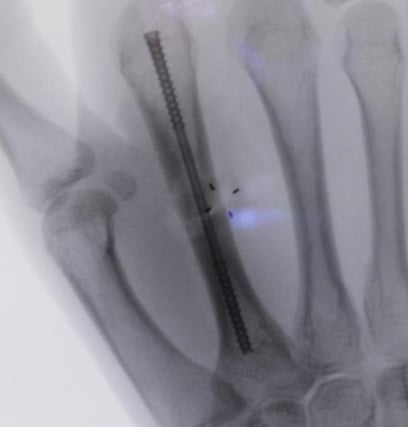
Below: Two week postoperative x-rays

Six week review
The surgical site had no lesions, discoloration or swelling (well healed incision with no signs or symptoms of infection). There was normal muscle bulk, no atrophy and muscle tone was normal.
The patient had full range of motion, with good grip strength and with no joint instability on provocative testing.
The patient was advised to continue increasing his activity as tolerated and within weight bearing limitations. The slow healing noted on the x-rays was discussed and the patient advised that a bone stimulator may be considered if delayed union continued to persist radiographically. The patient was advised to return in five weeks for a repeat examination and x-rays of the hand.
Below: Six week postoperative x-rays


Ten week review
The surgical site had no lesions, discoloration or swelling and the incision appeared well healed with no signs or symptoms of infection. Muscle bulk and tone were normal with no atrophy. The patient had full range of motion, with good grip strength and no joint instability on provocative testing.
The patient was advised to continue to increase his activity, no longer requiring weight bearing limitations. The slow healing observed on his x-rays was discussed however more healing was noted along the radial border of the osteotomy site when compared to the six week imaging. The patient was advised to return in four weeks for repeat examination and x-rays of the hand at which point a bone stimulator may be considered if delayed union persisted radiographically.
Below: 10 week postoperative x-rays


References
1. Field Orthopaedics. (2022). NX Nail Product Brochure. Brisbane, Australia: Field Orthopaedics.
2. Field Orthopaedics. (2023). NX Nail Long Surgical Technique. Brisbane, Australia: Field Orthopaedics.
FO-008357-MM Version 2 May 2024